Antimicrobial Evaluation of Phyllanthus amarus Leaf Extracts Against Beta-Lactamase Resistance Escherichia coli isolated from Eidolon helvum
-
Aladejana Oluwatoyin Modupe


Department of Biological Sciences, Microbiology Unit, Kings University, 220104, Odeomu, Osun, Nigeria
Akinmarin Boluwatife Morayo
Department of Chemical Sciences, Kings University, 220140, Odeomu, Osun, Nigeria
Ogidi Clement OlusolaDepartment of Biological Sciences, Microbiology Unit, Kings University, 220104, Odeomu, Osun, Nigeria
Oladele Johnson Olaleye
Department of Chemical Sciences, Kings University, 220140, Odeomu, Osun, Nigeria
Ogunlade Ayodele OluwayemisiDepartment of Food Technology, School of Science and Computer Studies, The Federal Polytechnic Ado Ekiti, Ado Ekiti, Ekiti, Nigeria
Olawoye AbimbolaDepartment of Biological Sciences, Microbiology Unit, Kings University, 220104, Odeomu, Osun, Nigeria
| Received 16 Feb, 2022 |
Accepted 09 May, 2023 |
Published 10 May, 2023 |
Background and Objective: Prophylactic use of medicinal plants in the treatment of human and animal infections and diseases has greatly improved over the years and continuously been explored. The study was conducted to assess the antimicrobial activity of Phyllanthus amarus against Escherichia coli isolated from Eidolon helvum (Straw Coloured Fruit Bat). Materials and Methods: A total of seventeen Escherichia coli isolates were grown on eosin methylene blue agar and MacConkey agar. Biochemical tests were carried out and antibiotic susceptibility tests were performed using Kirby-Bauer’s disc diffusion technique. Results: The results were interpreted according Clinical and Laboratory Standards Institute (CLSI). The aqueous and ethanol plant extracts of dry Phyllanthus amarus leaves were retrieved in ratio 3:1 of plant to water and plant to ethanol. The highest zones of inhibition recorded were 16 mm for aqueous extract and 14 mm for ethanol extract at concentration of 25 mg mL‾1. Bioactive compounds found in both aqueous and ethanol extracts of Phyllanthus amarus include alkaloids, saponins, flavonoids, anthraquinones and glycosides. Tannins were found in the aqueous extract only while terpenoids were found in the ethanol extracts only. Furthermore, the GC-MS analysis of the plant extract revealed that the aqueous and ethanol extracts are rich in phytochemicals which has been reported to have antioxidant and other biological activities. Conclusion: It was concluded that Phyllanthus amarus can be considered a good alternative to antibiotics in the case of infections and diseases caused by multi-drug resistant Escherichia coli from straw coloured fruit bats when taken in the appropriate dosage.
| Copyright © 2023 Modupe et al. This is an open-access article distributed under the Creative Commons Attribution License, which permits unrestricted use, distribution, and reproduction in any medium, provided the original work is properly cited. |
INTRODUCTION
The treatment of human and animal diseases and infections has greatly improved over the years due to the discovery of medicinal plants that grow in various environments. These medicinal plants have helped to make medicine available in local or rural areas where the healthcare system is not as developed as in urban areas. Some countries which do not have access to expensive technology and knowledge in producing drugs have also relied on the benefits of medicinal plants, especially those that do not require advanced processing techniques.
Phyllanthus amarus is a member of a large genus of flowering plants called Phyllanthus. It is commonly called ‘leaf flower’ because of its fruit, leaves and flowers appear fused. It is closely related to Phyllanthus niruri also called ‘gale of the wind’. There are about 800 species of the family of Euphorbiaceae commonly found in tropical and sub-tropical countries in the world1,2. Over 50% of all modern chemical drugs are of natural plant product origin and is essential in drug development programs of the pharmaceutical industry3,4. Phyllanthus amarus has been used in traditional medicine to cure health problems such as jaundice, dropsy, urinogenital disorders, kidney problems, diabetes and skin problems. It is also found to be effective against gastrointestinal disorders such as colic which is common in babies, diarrhoea, dysentery and constipation. In the case of wounds, the leaf is usually made into a paste and applied unto the wounded area1. The root extract of the plant is also useful in relieving stomach pain while the flower extract is applied externally as an antidote to snake bites5.
The ethanol and water extracts of Phyllanthus amarus have been observed to have antimicrobial effects against pathogenic bacteria such as Salmonella typhi. In another study the hexane, petroleum ether, chloroform, acetone and methanol extracts were found to be effective when tested against the organisms Klebsiella pneumonia, Proteus mirabilis, Streptococcus faecalis, Klebsiella pneumonia, Enterobacter species, Serratia marcescens, Staphylococcus aureus and Escherichia coli6. In an attempt to treat the E. coli infection complications emerged due to the bacterial resistance to most first-line antimicrobial agents7,8. Over the years, resistance to cephalosporins among members of the Enterobacteriaceae family has increased greatly because of the spreading of Extended-spectrum β-Lactamases (ESBL) that destroy beta-lactam antibiotics9,10.
Multiple antibiotic resistant E. coli isolated from straw coloured fruit bat is a threat to human health and most especially in areas where there is a little or no access to good medication. Therefore, this study aimed at investigating the antimicrobial efficacy of Phyllanthus amarus leaf extracts against beta-lactamase resistance Escherichia coli isolated from Eidolon helvum.
MATERIALS AND METHODS
Isolation and identification of the intestinal organism: A total of seventeen multiple antibiotics resistance Escherichia coli were gotten from the faecal samples of straw-coloured fruit bats from Ile-Ife, Osogbo and Ilesa, in Osun State Nigeria between October, 2018 and March, 2019. The research was carried out by the current rules and guidelines established for the care of laboratory animals in Kings University, Odeomu, Nigeria. The isolates were sub-cultured on MacConkey agar (LAB M, UK) to check for organism purity, from where suspensions were made in sterile normal.
Antibiotic susceptibility test: An appropriate quantity of Mueller-Hinton agar was weighed and dissolved in the appropriate volume of distilled water. The solution was autoclaved (Finlab Nigeria limited, Lagos, Nigeria) for 15 min at 121°C. A 24 hrs old culture was standardized and streaked evenly on each agar plate with a sterile swab stick and allowed to dry. The antibiotic discs (ThermoFisher Scientific, Waltham, MA, USA) were placed using a sterile pair of forceps. The plates were inverted and incubated (Finlab Nigeria limited, Lagos, Nigeria) at 37°C for 24 hrs.
Antibiotics used include, Amoxicillin (AMC), Ampicillin (AMP), Augmentin (AUG), Ceftazidime (CAZ), Cefuroxime (CRX), Ceftriaxone (CTR), Cloxacillin (CXC), Ertapenem (ETP), Erythromycin (ERY), Gentamicin (GEN) and Ofloxacin (OFL). Clear zone of inhibition around each disc was measured using a calibrated ruler in millimetre and interpreted using Clinical and Laboratory Standards Institute11.
The Multiple Antibiotic Index (MAR Index) for each of the isolates was calculated as indicated below:
where, a represents the aggregate antibiotic resistance score of all isolates, b is the number of antibiotics used and c is the number of isolates of the organism12.
Plant collection: The plant (20 g) was collected from Agunbelewo Area, Osogbo, Osun State Nigeria during the rainy season in June. This herb was identified and authenticated at the Department of Botany, Obafemi Awolowo University, Ile-Ife, Nigeria. The leaves and the beads were painstakingly picked from the front and thoroughly washed in clean water, then rinsed in distilled water. It was allowed to air dry at room temperature for several days until it was considered brittle enough for blending into fine powder.
Preparation of extracts: The powdered leaves were weighed and soaked in a ratio of 3:1 of plant to ethanol and plant to aqueous. The liquid was filtered and fix-dried. The extract (1.0 g) was dissolved in 10 mL of sterile distilled water to yield 100 mg mL–1. Therefore, 0.5, 0.25 and 0.125 g of plant extract weredissolved in 10 mL of sterile distilled water to yield 50 mg mL–1 concentration, 25 mg mL–1 concentration and 12.5 mg mL–1 concentration, respectively.
Susceptibility testing using the plant extract: This was done using the agar well diffusion method. Mueller-Hinton agar was prepared according to the manufacturer’s instructions11. Standardized isolates were streaked evenly on the agar plates and allowed to dry. Four wells were punched into the agar medium using a cork borer of diameter 0.4 mm and filled with 50, 25 and 12.5 mg mL–1 of aqueous plant extract, respectively. Another set was prepared in the same manner with ethanol plant extract. 2.5 mg mL–1 of standard antibiotic Ampiclox was used as a control in the fourth well. The plates were allowed to diffuse at room temperature and incubated at optimum temperature for 18-24 hrs. The plates were observed for zones of inhibition and Minimum Inhibitory Concentration (MIC).
Phytochemical screening and identification of bioactive compounds using GCMS: The following phytochemicals were screen for using standard methods saponins, tannins, phlobatannins, anthraquinones, alkaloids, flavonoids, alkaline reagent test, leadacetate test, evaluation of reducing sugars, terpenoids and glycosides. Gas Chromatography-Mass Spectrometry (GC-MS) analysis GC-MS analysis were carried out on a GC-MS-QP 2010 Plus Shimadzu system and Gas chromatograph interfaced to a mass spectrometer (GC-MS) instrument (BVK technology services, Boduppal, Hyderabad) employing the following conditions: Column Elite-1 fused silica capillary column (30m×0.25mm 1D×μL df, composed of 100% dimethyl polysiloxane). For GC-MS detection, an electron ionization system with ionization energy of 70 eV was used. Helium gas (99.999%) was used as the carrier gas at constant flow rate 1 mL min–1 and an injection volume of 2 μL was employed (Split ratio of 10:1) injector temperature -250°C, ion-source temperature 280°C. The oven (Finlab Nigeria limited, Lagos, Nigeria) temperature was programmed from 110°C (Isothermal for 2 min) with an increase of 10°C/min to 200°C then 5°C/min to 280°C/min, ending with a 9 min. isothermal at 280°C. Mass spectra were taken at 70 eV, a scan interval of 0.5 sec and fragments from 40 to 550 Da. Total GC running time was 36 min. The relative percentage amount of each component was calculated, by comparing its average peak area to the total areas. Software adopted to handle mass spectra and chromatogram was a turbomass. The detection employed the NIST Ver. 2.0 year 2009 library. Identification of components Interpretation on mass spectrum of GC-MS was done using the database of National institute of Standard and Technology (NIST) having more than 62,000 patterns. The mass spectrum of the unknown component was compared with the spectrum of the known components stored in the NIST library. The name, molecular weight and structure of the components of the test materials were ascertained13.
Statistical analysis: Results obtained were expressed as Mean±Standard Error of Mean (Mean±SD) and analysed using One-way Analysis of Variance (ANOVA) with the aid of SPSS 22.0 computer software package (SPSS Inc, Chicago, USA). Values at p<0.05 were considered significant.
RESULTS
The antibiotic susceptibility of the seventeen Escherichia coli were used in the study. The test results depicted that all the isolates were sensitive to gentamicin (GEN) and ofloxacin (OFL) (100%). However, the isolates showed (100% resistance to erythromycin (ERY), cloxacillin (CXC) and augmentin (AUG) (100%), while the resistance were also high to ceftazidime (CAZ) 88.2%, cefuroxime (CRX) 88.2%, amoxicillin (AMC) 70.6%, ETP) 70.6% and ampicillin (AMP) 47.1%, respectively as shown in Table 1.
Multiple antibiotic index (MAR) is a biological parameter to determine organisms with high risk of contamination where antibiotics are often used. The frequency of the multiple antibiotic resistance patterns of the E. coli isolates to various antibiotic and the Multiple Antibiotic Index (MAR Index) was as shown in Table 2. Escherichia coli isolates: E7B, E16A, E16B, E20 and E26A displayed the highest MAR index of 2.67 while E10, E22, E23 and E25 showed MAR index of 1.75. All the isolates showed a significant MAR index greater than 0.2 thus they are from high risk sources.
| Table 1: | Antibiotic susceptibility pattern of Escherichia coli isolates from faecal samples of straw coloured fruit bats in Osun State Nigeria | |||
Resistant isolates |
Susceptibility pattern |
||||
| Class of antibiotics | Antimicrobial agent (Abbreviation) |
Number |
Percentage |
Number |
Percentage |
| β-Lactam | AMC |
12 |
70.6 |
5 |
29 |
AMP |
8 |
47.1 |
9 |
53 |
|
AUG |
17 |
100 |
0 |
0 |
|
CXC |
17 |
100 |
0 |
0 |
|
| Cephalosporin | CAZ |
15 |
88.2 |
2 |
12 |
CRX |
15 |
88.2 |
2 |
12 |
|
CTR |
0 |
0 |
17 |
100 |
|
| Macrolide | ERY |
17 |
100 |
0 |
0 |
| Carbapenem | ETP |
12 |
70.6 |
5 |
29 |
| Aminoglycoside | GEN |
0 |
0 |
17 |
100 |
| Fluoroquinolone | OFL |
0 |
0 |
17 |
100 |
| AMC: Amoxicillin, AMP: Ampicillin, AUG: Augmentin, CAZ: Ceftazidime, CRX: Cefuroxime, CTR: Ceftriaxone, CXC: Cloxacillin, ETP: Ertapenem, ERY: Erythromycin, GEN: Gentamicin and OFL: Ofloxacin | |||||
| Table 2: | Multiple resistance patterns of Escherichia coli isolates from straw coloured fruit bat | |||
| Code | Resistance Pattern | Frequency |
MAR (%) n=17 |
MAR index |
| E1 | AUG, CAZ, CRX, CXC, ERY, ETP | 1 |
5.9 |
1.2 |
| E5, E26B | AMP, AUG, CAZ, CRX, CXC, ERY | 2 |
11.8 |
1.2 |
| E7A | AUG, CAZ, CRX, CXC, ERY | 1 |
5.9 |
0.83 |
| E7B, E16A, E16B, E20, E26A | AMC, AMP, AUG, CAZ, CRX, CXC, ERY, ETP | 5 |
29.4 |
2.67 |
| E9 | AMC, AUG, CXC, ERY, ETP | 1 |
5.9 |
0.83 |
| E10, E22, E23, E25 | AMC, AUG, CAZ, CRX, CXC, ERY, ETP | 4 |
23.5 |
1.75 |
| E11A | AUG, CRX, CXC, ERY | 1 |
5.9 |
0.57 |
| E11B | AMC, AMP, AUG, CAZ, CXC, ERY, ETP | 1 |
5.9 |
1.75 |
| E24 | AMC, AUG, CAZ, CRX, CXC, ERY | 1 |
5.9 |
1.2 |
The results of the Minimum Inhibitory Concentration (MIC) and zone of inhibition of aqueous and ethanol leaf extracts of Phyllanthus amarus on the E. coli isolates were shown in Table 3. At 25 mg mL–1, aqueous leaf extract of Phyllanthus amarus showed the highest zone of inhibition of 16 mm against E1. Nevertheless, the extract has no inhibition on isolate E5, E25 and E26. Similarly, ethanol leaf extract of Phyllanthus amarus showed the highest zone of inhibition of 14 mm against E10. However, the extract has no inhibition on isolate E5 as shown in Table 3.
The bioactive constituents and phytochemicals found in the aqueous and ethanol extracts of dry Phyllanthus amarus leaves were shown in Table 4. The result depicted that both extracts were rich in saponins, anthraquinones, alkaloids, flavonoids and glycosides.
| Table 3: | Zones of inhibition (mm) by Phyllanthus amarus extract at 25 mg mL–1 and minimum inhibitory concentration (mg mL–1) on beta-lactamase resistant E. coli isolated from straw coloured fruit bat | |||
| Code | Aqueous extract |
Ethanol extracts |
MIC aqueous extract |
MIC ethanol extract |
| E1 | 16 |
8 |
25 |
25 |
| E5 | 0 |
0 |
12.5 |
12.5 |
| E7A | 12 |
8 |
25 |
25 |
| E7B | 14 |
8 |
12.5 |
25 |
| E9 | 10 |
11 |
25 |
25 |
| E10 | 6 |
14 |
50 |
25 |
| E11A | 10 |
10 |
12.5 |
25 |
| E11B | 10 |
10 |
25 |
25 |
| E16A | 4 |
8 |
25 |
25 |
| E16B | 8 |
10 |
25 |
25 |
| E20 | 6 |
10 |
25 |
25 |
| E22 | 8 |
6 |
25 |
25 |
| E23 | 7 |
10 |
12.5 |
25 |
| E24 | 10 |
4 |
25 |
25 |
| E25 | 0 |
1 |
50 |
25 |
| E26 | 0 |
6 |
25 |
25 |
| Table 4: | Phytochemicals present in aqueous and ethanol extracts of Phyllanthus amarus | |||
| Class of Compound | Aqueous |
Ethanol |
| Saponins | + |
+ |
| Tannins | + |
- |
| Phlobatannins | - |
- |
| Anthraquinones | + |
+ |
| Alkaloids | + |
+ |
| Flavonoids | + |
+ |
| Terpenoids | - |
+ |
| Glycoside | + |
+ |
| Reducing Sugars | - |
- |
| +: Present and -: Absent | ||
| Table 5: | Phytochemicals present in aqueous extract | |||
| RT | Phytochemicals | Composition (%) |
| 2.629 | 1-propanol, 2-methyl- | 1.26 |
| 2.646 | 2-propenal | 90.22 |
| 4.066 | Acetic acid, butyl ester | 2.32 |
| 4.341 | Ethanamine, N-pentylidene- | 0.39 |
| 4.629 | Ethylbenzene | 0.22 |
| 4.82 | n-Butyl ether | 1.24 |
| 4.877 | 2,2-Dimethyl-1,2-butanediol | 0.01 |
| 6.39 | 1,2-Dimethylasiridine | 0.04 |
| 6.567 | Propan-2-one o-(2-chloro-1-methyl-ethyl)-oxime | 0 |
| RT: Retention time | ||
| Table 6: | Phytochemicals present in ethanol extract | |||
| RT | Phytochemicals | Composition (%) |
| 2.226 | Ethyl acetate | 0.1 |
| 2.628 | Diethanolamine | 0.18 |
| 2.905 | 2-propenal | 29.16 |
| 4.06 | Acetic acid, butyl ester | 0.31 |
| 4.341 | Propanoic acid, 2-methyl-, 2,2-dimethyl-1-(2-hydroxy-1-methylethyl)propyl ester | 0.05 |
| 4.629 | Ethylbenzene | 0.04 |
| 4.707 | p-xylene | 0.02 |
| 4.82 | n-Butyl ether | 0.11 |
| 6.961 | Ethoxy (methoxy) methylsilane | 0.07 |
| RT: Retention time | ||
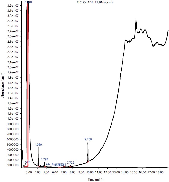
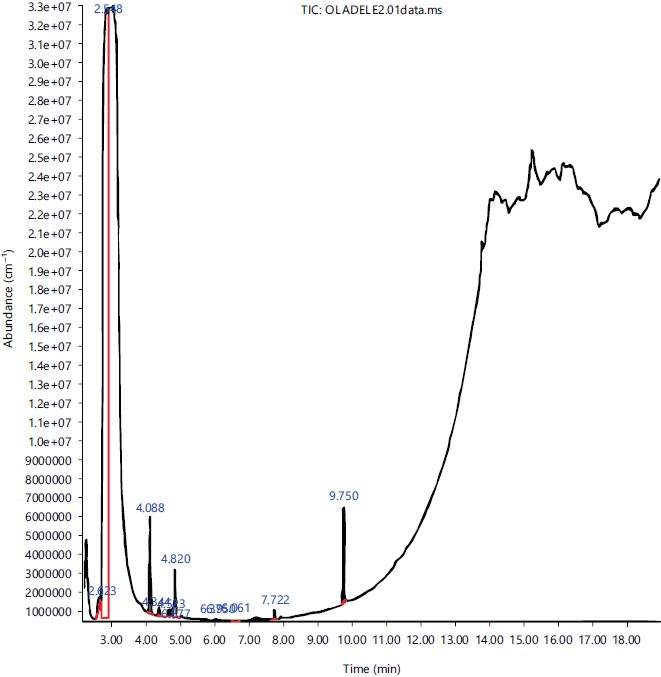
The GC-MS Chromatogram of aqueous and ethanol extract of leaves of P. amarus, respectively. While Table 5 and 6 showed that the bioactive compounds present in the aqueous and ethanol extract of leaves of P. amarus, respectively. The most abundant phytochemical in both extracts was 2-propenal as shown in Fig. 1 and 2.
|
|
DISCUSSION
The results obtained in this research showed that both the aqueous and the ethanolic extracts of the leaves have antibacterial activity on Escherichia coli, which was in an agreement with the findings of Silva et al. 15. The isolates were susceptible to both the aqueous (E1) and ethanolic (E2) plant extracts and the Minimum Inhibitory Concentration (MIC) was observed to be 25 mg mL–1 with only two isolates (11.8%) showing resistance to all concentrations of the extracts. However, in this research, the antimicrobial effects of the aqueous extracts proved to be more effective than the ethanolic extracts. In the research carried out by Natarajan et al.16, various extracts used were found to be effective against E. coli.
Previous studies and research have proved that Phyllanthus amarus is useful in medicine. However, this particular study is unique due to its focus on the antimicrobial effect of the leaf extracts of Phyllanthus amarus against E. coli which is commonly responsible for gastrointestinal disease and urinary tract infections. Saeidi et al.14 revealed the antibacterial activity of extracts of the root and leaves on Phyllanthus amarus against ESBL-producing Escherichia coli and he emphasized the importance of finding compounds that have potential antimicrobial activity against ESBL organisms due to the rising antibiotic resistance to drug classes such as ß-lactams.
The antibacterial activities of most plant extracts are dependent on their concentration. In this study the antimicrobial activities also increase as the concentration increases, therefore the most active concentration was seen at the highest concentration used. This also was in agreement with the study of Hafidh et al.17, which shows that the zone of growth inhibition increased with an increase in the concentration of the extracts. However, the more effective a low concentration of plant extract is the better, it is to be considered as an antimicrobial agent against Escherichia coli and other bacteria.
Preliminary phytochemical screening of the plant extracts revealed the presence of bioactive compounds in the leaves of Phyllanthus amarus such as saponins, anthraquinones, alkaloids, flavonoids, tannins and others which correlates with the reports of Akinjogunla et al.14. Saponins have anti-hyper cholesterol and cardiac depressant properties which are believed to contribute to the treatment of hypertension in patients18. Glycosides were also present and they are used in the treatment of heart diseases. Alkaloids and flavonoids are noted as antimicrobial, antioxidants and anti-inflammatory agents19.
The extracts of the root and leaf of Phyllanthus amarus has been assessed against extended spectrum lactamase (ESBL) producing Escherichia coli isolated from the stool samples of HIV seropositive patients and have proven susceptible at different concentrations14. In another study, the antimicrobial activity of Phyllanthus amarus was tested on the normal flora of the human intestine and it was effective. Although it was advised that the herbal infusions are taken with caution and supervision15.
The GC-MS analysis showed the existence of various compounds with different chemical structures. The presence of various bioactive compounds confirms the application of P. amarus for various ailments by traditional practitioners. However, the isolation of individual phytochemical constituents may proceed to find a novel drug compound. This study is limited to aqueous and methanol leaf extracts of Phyllanthus amarus against multidrug resistance E. coli. Each isolated compound from the extracts can been further explore to elucidate the antimicrobial principles of the plant. Also, other multidrug resistant bacterial strains could be further investigated to potentiate the antimicrobial potency of phytochemicals from extracts of Phyllanthus amarus. This study recommended that further studies and research should be channelled towards local herbal plant extracts and their antimicrobial activity to produce alternative medicine that would be available to patients in poor communities or the case of unavailability of antibiotics.
CONCLUSION
Phyllanthus amarus leaf extract inhibits bacterial growth and can be used as an alternative medicine in the case of multi-drug resistance to antibiotics of virulent E. coli strains that cause diseases. It is also beneficial in situations where commercial antibiotics are not available or the patient has developed resistance to the prescribed antibiotic medication. This can be achieved by appropriate animal and human studies and testing in order to create a standardized dose that does not cause complications to the body system.
SIGNIFICANCE STATEMENT
This study discovered that the phytochemicals obtained from Phyllanthus amarus leaf extract can be beneficial for the treatment and/or management of multi-drug resistance infections, specifically those that are caused by Escherichia coli e.g., ulcerative colitis. This study will help researchers to uncover the critical areas in the pathogenesis and treatment of multi-drug resistance infections that many researchers were not able to explore. Thus suggesting Phyllanthus amarus leaf extract is a potential drug candidate for the treatment of infections caused by Escherichia coli.
REFERENCES
- Joseph, B. and S.J. Raj, 2011. An overview: Pharmacognostic properties of Phyllanthus amarus Linn. Int. J. Pharmacol., 7: 40-45.
- Oladele, J.O., E.I. Ajayi, O.M. Oyeleke and O.T. Oladele et al., 2020. A systematic review on COVID-19 pandemic with special emphasis on curative potentials of Nigeria based medicinal plants. Heliyon, 6: e04897.
- Oladele, J.O., O.M. Oyeleke, O.T. Oladele and A.T. Oladiji, 2021. COVID-19 treatment: Investigation on the phytochemical constituents of Vernonia amygdalina as potential coronavirus-2 inhibitors. Comput. Toxicol., 18: 100161.
- Eweka, A.O. and A. Enogieru, 2011. Effects of oral administration of Phyllanthus amarus leaf extract on the kidneys of adult Wistar rats-A histological study. Afr. J. Tradit. Complementary Altern. Med., 8: 307-311.
- Patel, J.R., P. Tripathi, V. Sharma, N.S. Chauhan and V.K. Dixit, 2011. Phyllanthus amarus: Ethnomedicinal uses, phytochemistry and pharmacology: A review. J. Ethnopharmacol. Pharmacol., 138: 286-313.
- Ribeiro, A.M.B., J.N. de Sousa, L.M. Costa, F.A. de Alcântara Oliveira and R.C. dos Santos et al., 2019. Antimicrobial activity of Phyllanthus amarus Schumach. & Thonn and inhibition of the NorA efflux pump of Staphylococcus aureus by Phyllanthin. Microb. Pathogen., 130: 242-246.
- Sabaté, M., G. Prats, E. Moreno, E. Ballesté, A.R. Blanch and A. Andreu, 2008. Virulence and antimicrobial resistance profiles among Escherichia coli strains isolated from human and animal wastewater. Res. Microbiol., 159: 288-293.
- Aladejana, O.M., A.O. Oluduro, O.A. Thonda, A.O. Ogunlade and O. Famurewa, 2022. Molecular characterization of resistance and virulence genes in Escherichia coli isolated from bats (Eidolon helvum) faeces in Osun State, Nigeria. J. Adv. Microbiol., 22: 37-48.
- Shaikh, S., J. Fatima, S. Shakil, S.M.D. Rizvi and M.A. Kamal, 2015. Antibiotic resistance and extended spectrum beta-lactamases: Types, epidemiology and treatment. Saudi J. Biol. Sci., 22: 90-101.
- Aladejana, O.M. and A.O. Olufunke, 2021. Molecular characterisation of extended spectrum β-lactamase-producing- enterobacteriaceae from bats faeces in Osun State, Nigeria. Biol. Nyssana, 12: 79-86.
- Oludare, T.O., M. Agbi, O.A. Thonda and O.M. Aladejana, 2019. Pre/post plasmid curing and killing kinetic reactivity of Discorea bulbifera Linn against multiple antibiotics resistant clinical isolates, using Escherichia coli as a case study. Int. J. Cell Sci. Mol. Biol., 6: 46-56.
- Aladejana, O.M., J.O. Oluyege, T.O. Olowomofe, I.E. Obayemi and D.E. Oluyege, 2021. Multiple antibiotic resistance of airborne bacteria in outdoor markets in Ado-Ekiti Metropolis. Serb. J. Exp. Clin. Res., 2021.
- Oladele, J.O., J.C. Anyim, O.M. Oyeleke, B.D. Olowookere, M.O. Bamigboye, O.T. Oladele and A.T. Oladiji, 2021. Telfairia occidentalis mitigates dextran sodium sulfate‐induced ulcerative colitis in rats via suppression of oxidative stress, lipid peroxidation, and inflammation. J. Food Biochem., 45:e13873.
- Saeidi, S., N.A. Boroujeni, H. Ahmadi and M. Hassanshahian, 2015. Antibacterial activity of some plant extracts against extended-spectrum beta-lactamase producing Escherichia coli isolates. Jundishapur J. Microbiol., 8: e15434.
- Silva, E., S. Fernandes, E. Bacelar and A. Sampaio, 2016. Antimicrobial activity of aqueous, ethanolic and methanolic leaf extracts from Acacia Spp. and Eucalyptus nicholii. Afr. J. Tradit. Complementary Altern. Med., 13: 130-134.
- Natarajan, D., R. Srinivasan and M.S. Shivakumar, 2014. Phyllanthus wightianus Mull. Arg.: A potential source for natural antimicrobial agents. BioMed Res. Int., 2014: 135082.
- Hafidh, R.R., A.S. Abdulamir, L.S. Vern, F. Abu Bakar, F. Abas, F. Jahanshiri and Z. Sekawi, 2011. Inhibition of growth of highly resistant bacterial and fungal pathogens by a natural product. Open Microbiol. J., 5: 96-106.
- Singh, D. and P.K. Chaudhuri, 2018. Structural characteristics, bioavailability and cardioprotective potential of saponins. Integr. Med. Res., 7: 33-43.
- Tungmunnithum, D., A. Thongboonyou, A. Pholboon and A. Yangsabai, 2018. Flavonoids and other phenolic compounds from medicinal plants for pharmaceutical and medical aspects: An overview. Medicines, 5: 93.
How to Cite this paper?
APA-7 Style
Modupe,
A.O., Morayo,
A.B., Olusola,
O.C., Olaleye,
O.J., Oluwayemisi,
O.A., Abimbola,
O. (2023). Antimicrobial Evaluation of Phyllanthus amarus Leaf Extracts Against Beta-Lactamase Resistance Escherichia coli isolated from Eidolon helvum. Research Journal of Microbiology, 18(1), 1-10. https://doi.org/10.3923/rjm.2023.01.10
ACS Style
Modupe,
A.O.; Morayo,
A.B.; Olusola,
O.C.; Olaleye,
O.J.; Oluwayemisi,
O.A.; Abimbola,
O. Antimicrobial Evaluation of Phyllanthus amarus Leaf Extracts Against Beta-Lactamase Resistance Escherichia coli isolated from Eidolon helvum. Res. J. Microbiol 2023, 18, 1-10. https://doi.org/10.3923/rjm.2023.01.10
AMA Style
Modupe
AO, Morayo
AB, Olusola
OC, Olaleye
OJ, Oluwayemisi
OA, Abimbola
O. Antimicrobial Evaluation of Phyllanthus amarus Leaf Extracts Against Beta-Lactamase Resistance Escherichia coli isolated from Eidolon helvum. Research Journal of Microbiology. 2023; 18(1): 1-10. https://doi.org/10.3923/rjm.2023.01.10
Chicago/Turabian Style
Modupe, Aladejana, Oluwatoyin, Akinmarin Boluwatife Morayo, Ogidi Clement Olusola, Oladele Johnson Olaleye, Ogunlade Ayodele Oluwayemisi, and Olawoye Abimbola.
2023. "Antimicrobial Evaluation of Phyllanthus amarus Leaf Extracts Against Beta-Lactamase Resistance Escherichia coli isolated from Eidolon helvum" Research Journal of Microbiology 18, no. 1: 1-10. https://doi.org/10.3923/rjm.2023.01.10

This work is licensed under a Creative Commons Attribution 4.0 International License.